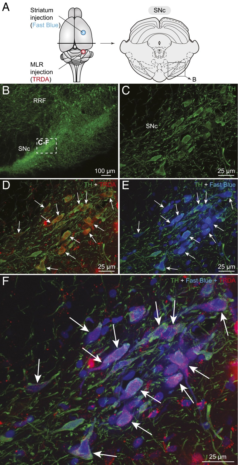
Fig. 5.
TH neurons projecting to the striatum and to the MLR are intermingled in the SNc. (A) Schematic dorsal view of the rat brain with the approximate location of the tracer injection sites in the striatum (Fast blue, blue) and in the pedunculopontine nucleus (TRDA, red). (Right) Diagram illustrating a cross-section at the level of the SNc with the approximate location of the photomicrographs shown in B. (B and C) DA neurons of the SNc were identified using immunofluorescence against TH (green). (B) A magnification of the rectangle in A. (D) Injection of a tracer in the MLR- (TRDA, red) (Fig. S7 C and D) labeled numerous TH+ cells (green) in the SNc. (E) Injection of another tracer in the striatum (Fast blue, blue) (Fig. S7 A and B) also labeled TH+ cells (green) in the SNc. (F) The photomicrographs were merged to show the three markers. In D–F; the arrows indicate numerous examples of TH+ cells of the SNc that project to the MLR and to the striatum. (C–F) Magnifications of the dashed rectangle in B.

