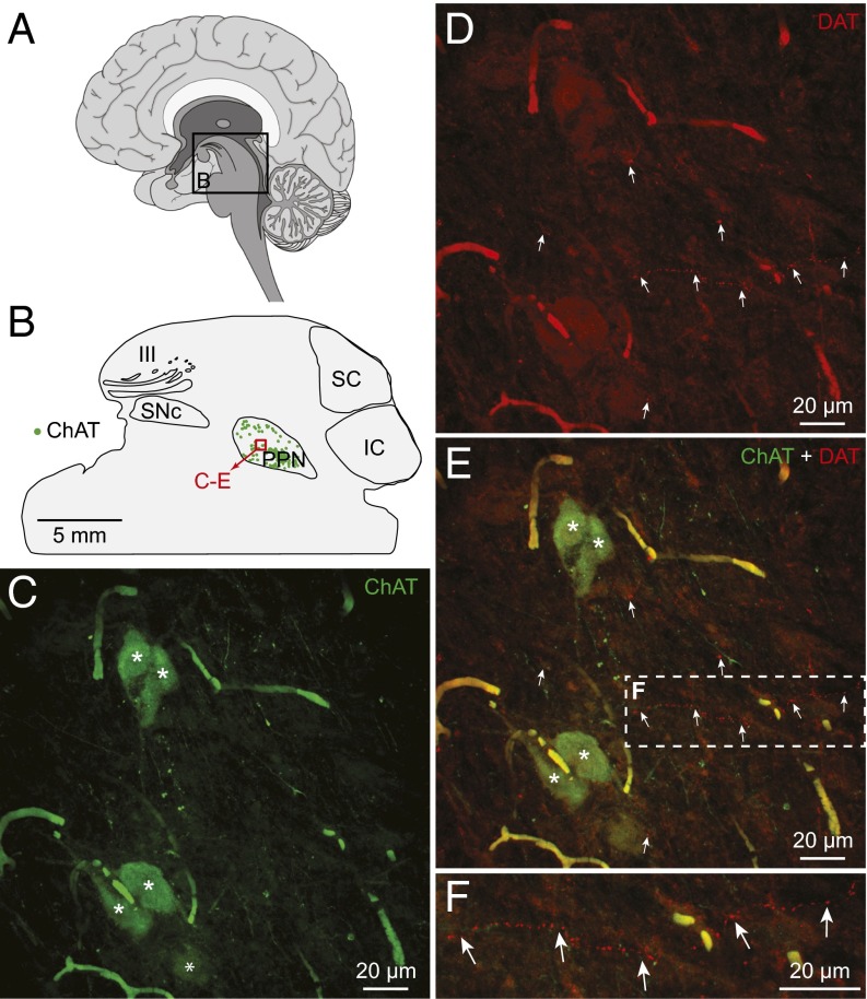
Fig. 7.
Dopaminergic innervation of the MLR in the human brain. (A) Schematic sagittal view of a human brain illustrating the approximate location of the brain section at the level of the PPN, considered part of the MLR (35). (B) Schematic sagittal view of the brain section (laterality: 5.8 mm from midline) used for the illustrated immunofluorescence experiment. The location of PPN cells positive for ChAT (green) is illustrated. The approximate location of the photomicrographs in C–F is shown by a red square. (C–F) Fibers and varicosities containing the DAT (red, highlighted by arrows) in proximity with cholinergic cells (ChAT, green) in the PPN. (F) A magnification of the dashed rectangle in E. The images in C–F were obtained by projecting on the same plane a stack of 20 images (0.96-µm deepness per image) acquired along the z axis. Asterisks illustrate cholinergic cells bodies.

