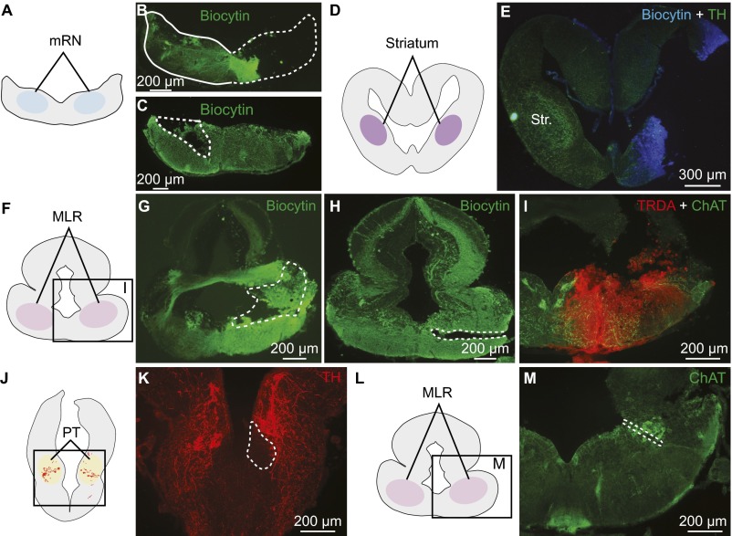
Fig. S2.
Histological controls for injection, stimulation, and recording sites in salamanders. (A–C) Typical injection sites in the mRN (enclosed by white dashed line) of the retrograde tracer biocytin (green). The injection site corresponds to the location of cellular bodies and dendrites of mRN reticulospinal neurons (7). (D and E) Typical injection site in the striatum (Str.) of the tracer (biocytin, blue) used to retrogradely label neurons in the PT. The striatum can be identified on the intact side, where its innervation by TH+ fibers is clearly visible. (F–I) Typical injection sites in the MLR of the retrograde tracers biocytin (G and H, enclosed by white dashed line) or TRDA (I), both used to retrogradely label PT neurons. The injection site coincides with the MLR cells positive for ChAT neuronal population of the laterodorsal tegmental nucleus (Fig. 1). (J and K) For voltammetry and calcium imaging experiments, the PT stimulation site (enclosed by white dashed line) was identified either by an electrolytic lesion or by the lesion made with the microinjection pipette used for chemical stimulation. The stimulation site coincided with the location of TH+ neurons (red). (L and M) The voltammetry recording site in the MLR was identified by electrolytic lesion (enclosed by white dashed line in M). The recording site coincides with the location of the cholinergic cells of the MLR (green), where a large number of dopaminergic fibers was observed (Fig. 1 A–G and Fig. S1 A–G).

