Fig. S5.
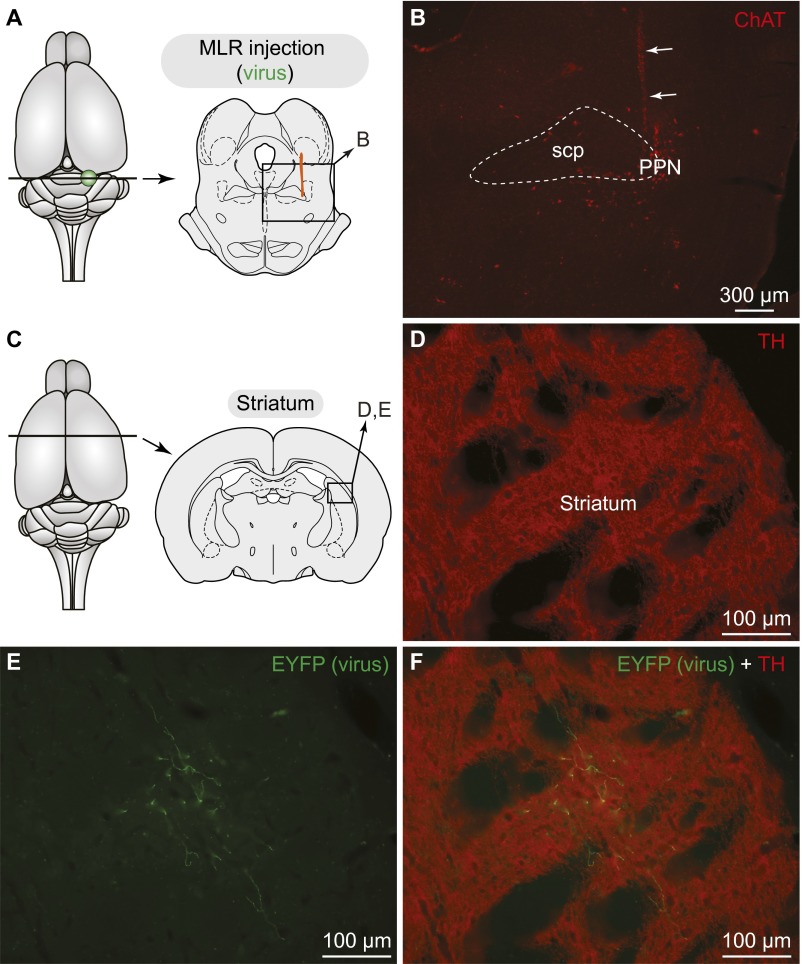
Fibers labeled in the striatum following injection of a Cre-dependent virus in the MLR of TH-Cre+ rats. (A) Schematic dorsal view of the rat brain. (Right) Diagram illustrating a cross-section at the level of the MLR with the approximate location of the photomicrograph shown in B. (B) Virus injection site in the cluster of cholinergic cells (ChAT+, red) of the PPN, an anatomical landmark of the MLR in rats (2). The trajectory of the injection needle is highlighted with arrows. This was used as a landmark, because the expression of the EYFP encoded by the virus could not be detected in this set of experiments at the level of the injection site in the PPN. The superior cerebellar peduncle (scp) is delineated with thin dashed lines. (C) Schematic dorsal view of the rat brain. (Right) Diagram illustrating a cross-section at the level of the striatum with the approximate location of the photomicrographs shown in D–F. (D) TH+ (red) innervation of the striatum. (E) Fibers in the striatum labeled following PPN injection of a Cre-dependent virus encoding for the EYFP (green). (F) The photographs were merged to see the two markers. Data from A–F were obtained from the same animal.

