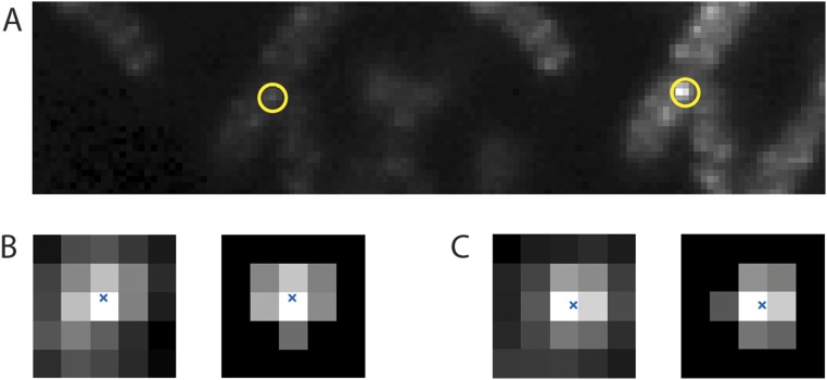
Fig. S1.
(A) An image of 128 × 32 pixels averaged over 199 frames taken before and after the bleaching pulse showing five cells with about 12 fluorescent motors excited in the S, with one motor circled in yellow. The P fluorescence is shown on the left, and the S fluorescence is shown on the right. (B, Left) Pixels in the vicinity of the motor image in P (B, Right) cropped to within 150 nm of that image’s center of mass. The image’s center of mass is marked with a blue x. (C) Pixels in the vicinity of the motor image in S displayed in a similar manner.

