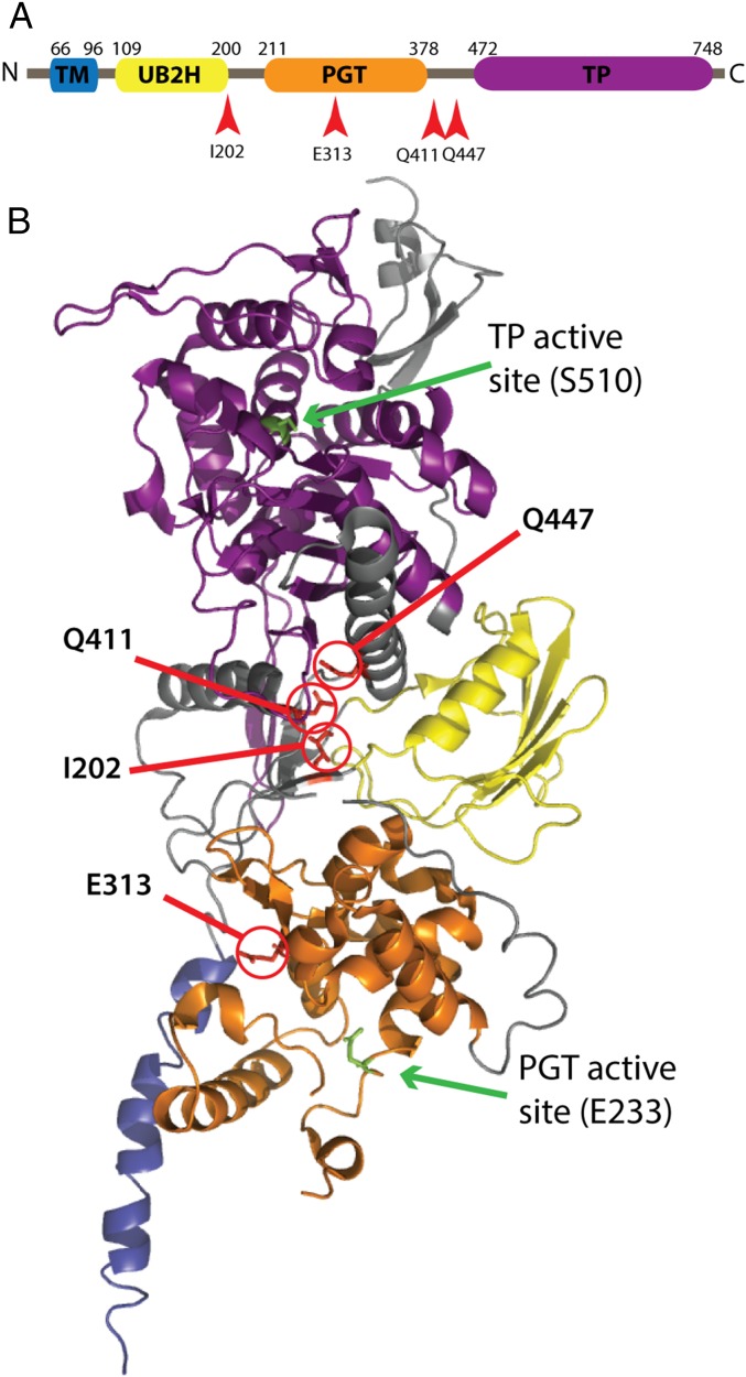
Fig. 1.
Location of altered residues in PBP1b* variants. (A) Schematic of the PBP1b primary structure: transmembrane helix (TM), UB2H domain, peptidoglycan glycosyltransferase (PGT), and transpeptidase (TP). (B) Crystal structure of PBP1b (PDB: 3FWM) (19) with the locations of the altered residues in the PBP1b* variants highlighted in red, along with the catalytic residues for both active sites in green. Domains in the structure are colored to match those in A.

