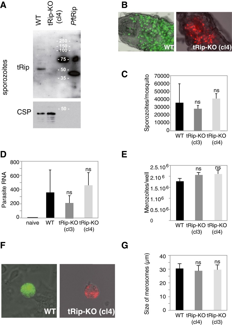
Fig. S7.
Prepatent stages of the tRip-KO parasite. (A) Western blot of purified WT and tRip-KO sporozoites. (B) Oocysts of the WT (Left) or tRip-KO (cl4; Right) clones developing in the midgut of A. stephensi mosquitoes 11 d after the mosquito’s infective blood meal. (C) Mosquitoes infected with WT or tRip-KO (cl3 and cl4) parasites harbored similar numbers of sporozoites in their salivary glands 18 d (and onward) after infection. Means are from two independent experiments, with n = 16–30 female mosquitoes. (D) Parasite loads in the liver 46 h after i.v. injection of 104 sporozoites were quantified by RT-PCR analysis. The histogram shows the abundance of parasite RNA obtained from 16 infected mice (for each clone), using the mouse HPRT gene as a standard. (E) Incubation of 2 × 10⁵ HepG2 hepatoma cells with 5 × 10⁴ salivary gland WT or tRip-KO (cl3 and cl4) sporozoites (per well) generated similar numbers of merozoites. (F) Representative WT (Left) and mutant (Right) merosomes, which are merozoite-filled vesicles that bud off from infected HepG2 cells after 60–65 h of liver stage development. (G) Size of merosomes released by WT and tRip-KO liver stages. Error bars are SDs, unpaired t test: ns indicates that values are not significantly different (P > 0.1).

