
Figure 3.
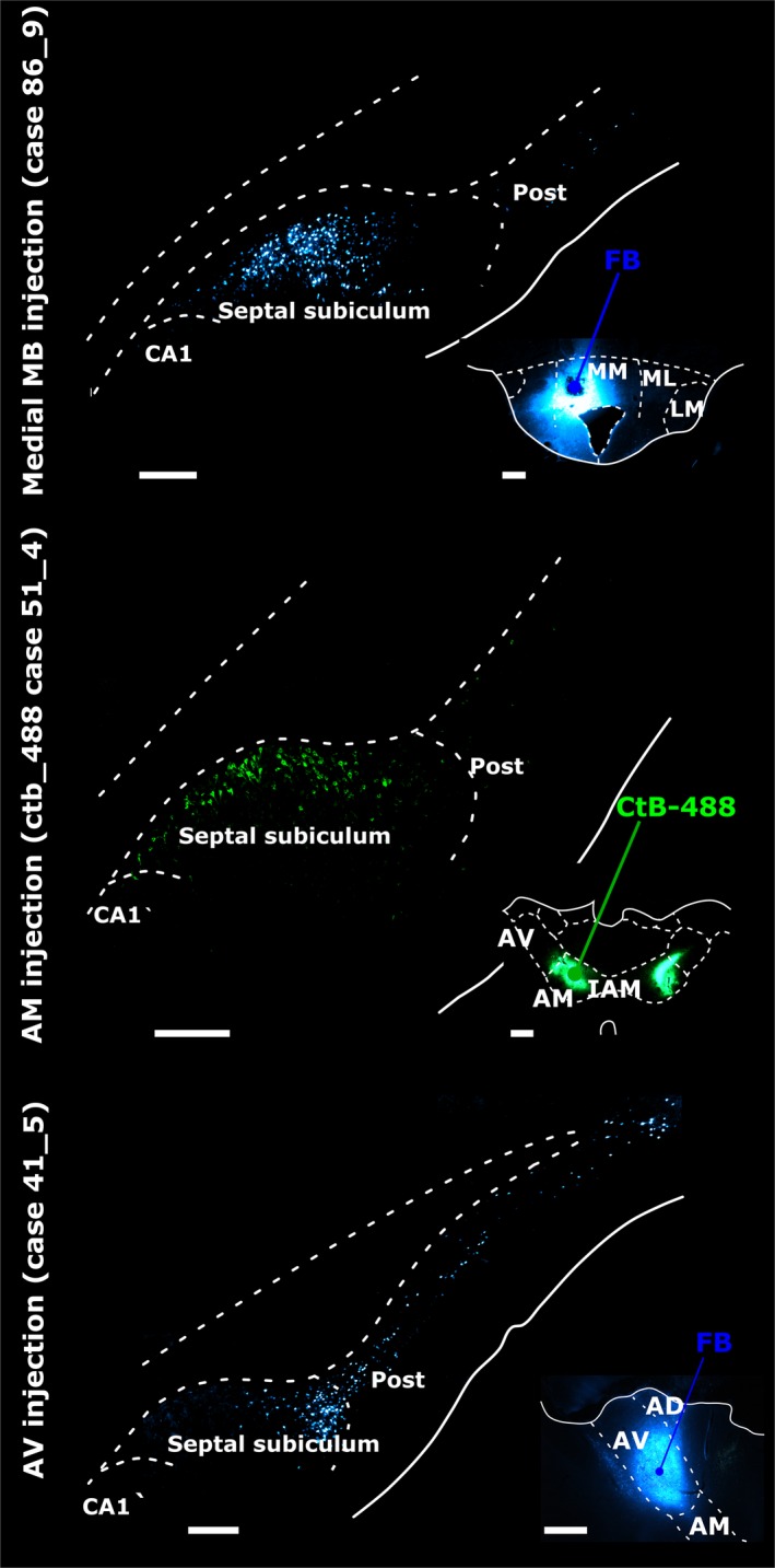
Retrograde neuronal label in the septal (dorsal) subiculum, postsubiculum (Post) and CA1 in the left hemisphere following tracer injections into (top panel) pars medialis of the medial mammillary nucleus (medial MB), (middle panel) the AM and (bottom panel) the AV. In each case, insets show injection site locations and tracer spread. Injections into the medial mammillary nucleus resulted in most labelled cells in mid‐subiculum regions R2 and R3 (top panel). Subiculum label density was greater proximal to CA1 following AM injections (middle panel), while tracer injections into AV resulted in most retrogradely label distal to CA1 (bottom panel). AD, anterodorsal nucleus; CA1, CA1 hippocampal subfield; CtB‐488, non‐toxic B‐subunit of cholera toxin, Alexa‐Fluor 488 conjugate; IAM, interanteromedial nucleus; LM, lateral MB nucleus; ML, pars lateralis of the medial mammillary nucleus; MM, pars medialis of the medial mammillary nucleus. Scale bars, 500 μm (injection sites), 250 μm (subiculum).