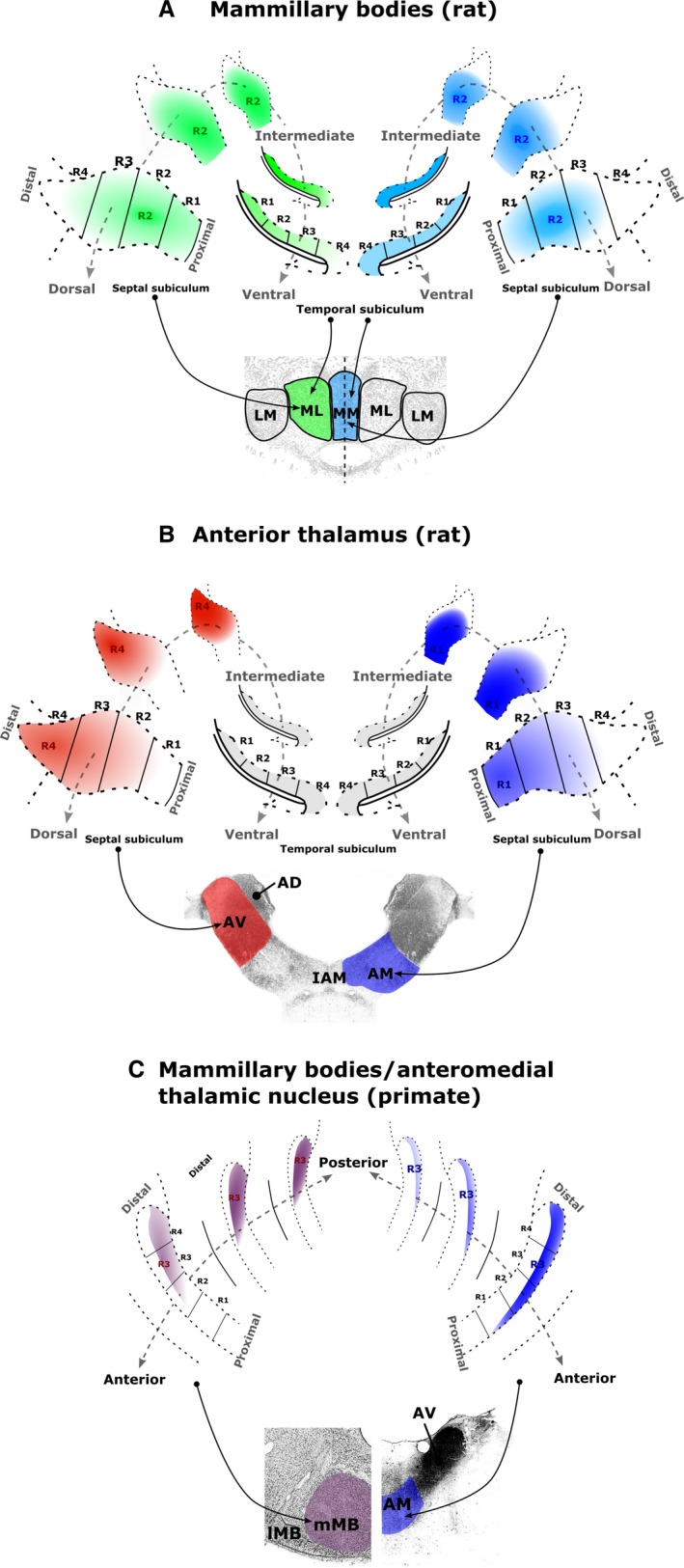
Figure 10.

Schematic summaries of the topographic sources of the subicular inputs to (A) the rat medial MBs, (B) the rat anterior thalamic nuclei and (C) the macaque medial MBs (left) and AM (right). Denser sources of subicular label correspond to denser colours. (A) Subicular inputs to the rat medial mammillary pars lateralis (green) and pars medialis (blue) arise predominantly from regions R1–3. For both mammillary sub‐nuclei, the proportion of subiculum‐projecting cells increases approaching the intermediate hippocampus. (B) Inputs to the rat AV (red) arise predominantly from distal regions (R3–4) of the septal–intermediate subiculum, while the corresponding inputs to the rat AM (blue) mainly arise from the proximal regions (R1–2) of the septal–intermediate subiculum. (C) Subicular cells that project to the macaque medial MBs (left) are more numerous in the posterior hippocampus while subicular cells that project to the AM are more numerous in the anterior hippocampus. The two sets of inputs predominantly arise from the distal subiculum (especially R3) but from different laminae. AD, anterodorsal thalamic nucleus; LM, lateral mammillary nucleus; ML, medial mammillary nucleus, pars lateralis; MM, medial mammillary nucleus, pars medialis.
