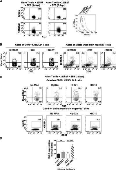
Figure 4.

Binding of KIR‐3DL2 to B27 heavy chains promotes the survival of activated naive CD4+ T cells. A, Left, Flow cytometry plots show staining for KIR‐3DL2 or IgG2a isotype control in CD45RO+ T cells from a patient with AS after 5 days of stimulation with LBL.721.220 cells (transfected with HLA–B7 [220B7] or HLA–B27 [220B27]) and staphylococcal enterotoxin B (SEB). Right, KIR‐3DL2 expression in cells under each condition is shown as the percentage of maximum (Max). B, OX40+KIR‐3DL2+ naive CD4+ T cells from a healthy control were analyzed by Live/Dead staining to exclude dead cells (left) and identify viable cells (right), after 5 days of stimulation with the indicated LBL.721.220 cells and SEB. Nontransfected LBL.721.220 cells (220) were used as a transfection control. Isotype control monoclonal antibody (mAb) did not stain (results not shown). C, CD69+KIR‐3DL2+ T cells among naive CD4+ T cells from a healthy control were analyzed by Live/Dead staining to exclude dead cells (top) and identify viable cells (bottom), after 2 days of stimulation with LBL.721.220 B27+ cells and SEB with or without the indicated mAb. In the flow cytometry analyses, representative results from 1 of 3 independent experiments are shown. D, Bcl‐2 expression was assessed by quantitative polymerase chain reaction in healthy control naive CD4+ T cells cocultured with the indicated LBL.721.221 cells and SEB for 6 or 24 hours. Results are the mean ± SEM of 5 independent experiments. P values were determined by analysis of variance. NS = not significant (see Figure 1 for other definitions).
