Figure 1.
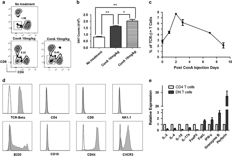
The proportion of CD3+CD4−CD8− double-negative T cells (DNT) was upregulated following ConA administration in C57BL/6 mice. DNT were significantly induced by ConA stimulation in a dose- ((a and b), n=3 in each group) and time- (c) dependent manner (n=4). The induced DNT (Gray histograms) highly expressed αβTCR, B220, CD44 and CXCR3, whereas no expression of CD4, CD8, CD19 and NK1.1 was observed. Mouse isotype Ig used as control (white histograms, d). Gene expression profile of ConA-induced DNT (e). **P<0.01, *P<0.05

