Figure 5.
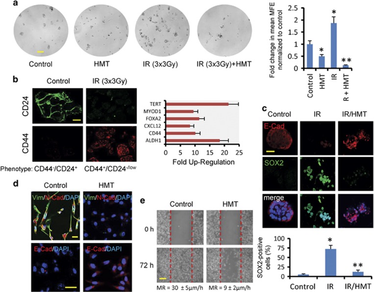
(a) The effects of fractionated radiation on secondary mammosphere-forming efficiency (MFE) in MCF7 cells. Primary MCF7 mammospheres were plated at single-cell densities and irradiated (3 Gy on three consecutive days) in the absence or presence of the HMT. Four days later, MFE was calculated. Scale bar refers 100 μm. *P<0.05 compared with the control mammospheres. **P<0.05 compared with the irradiated mammospheres. (b) IR induces stemness in survivor cells. MCF cells were irradiated (5 Gy), and, after 72 h, the surviving cells were cultured in adherent plates. At 5 days after replating, the cells were analyzed using immunofluorescence for CD24 and CD44, and the expression levels of 84 genes related to the identification, growth and differentiation of stem cells were analyzed using PCR arrays. In comparison with resting MCF7 cells, IR-survivor cells showed elevated levels of some recognized stemness markers (P<0.001). Scale bar refers 40 μm. (c) IR at 10 Gy accelerated dedifferentiation in MCF7-derived mammospheres. After 3 days, the morphological changes in the IR-treated primary mammospheres were paralleled by a decrease in E-cadherin (red) and the enrichment of SOX2 (green), as determined using confocal microscopy. However, the HMT inhibited the dedifferentiation of IR-treated primary mammospheres. Scale bars refers 20 μm. The number of SOX2-positive cells was expressed as a percentage after 200 cells were counted in different microscopic fields. The bars indicated±S.D. from three independent experiments. *P<0.05 compared with the control mammospheres. **P<0.05 compared with both the control and the irradiated mammospheres. (d) The expression levels of vimentin (green), N-cadherin (red) and E-cadherin (red) were analyzed using immunofluorescence staining in MDA-MB-231 cells before and after 72 h of treatment with the HMT. Scale bar refers to 20 μm. (e) Migration was analyzed in the control MDA-MB-231 cells and the cells treated with the HMT using wound-healing assays. A representative experiment is shown, and the statistical analysis for the migration rate (MR) is described (mean±S.D. from five independent experiments; P<0.002). Scale bar refers to 100 μm

