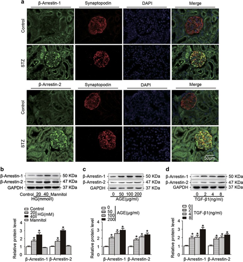
Figure 3.
β-Arrestin-1 and β-arrestin-2 were upregulated in podocytes under hyperglycemia both in vivo and in vitro. (a) Representative confocal microscopic images showing the upregulation of podocyte β-arrestin-1/2 in the kidney from STZ-induced diabetic mice; synaptopodin was used as a podocyte marker. (b) Representative western blotting gel documents and summarized data showing the protein levels of β-arrestin-1/2 in podocytes treated with HG (final concentration 20 or 40 mmol/l in medium) for 24 h. (c) Representative western blotting gel documents and summarized data showing the protein levels of β-arrestin-1/2 in podocytes with AGE (50–200 μg/ml) for 24 h. (d) Representative western blotting gel documents and summarized data showing the protein levels of β-arrestin-1/2 in podocytes treated with TGF-β1 (2–8 ng/ml) for 24 h. (means±S.E.M.; n=6; *P<0.05 versus control)

