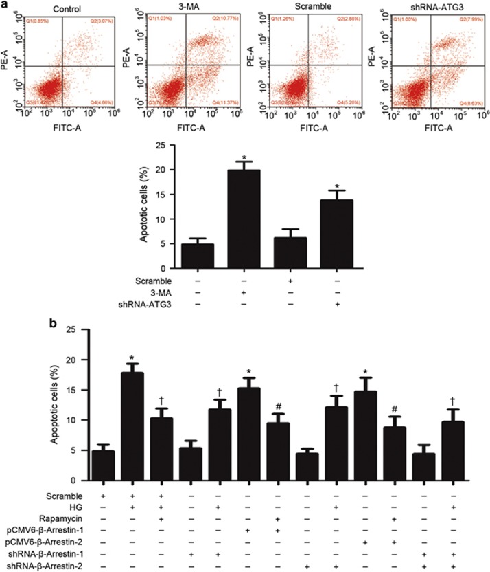
Figure 5.
Autophagy inhibition by β-arrestins reduced podocyte viability by flow cytometric analysis. (a) Autophagy inhibition by gene silencing of ATG-3- or 3-methyladenine (3-MA)-induced apoptosis. (b) HG-induced apoptosis was alleviated by gene silencing of β-arrestins as well as by restoring defective autophagy with low dose of rapamycin. Overexpression of β-arrestins by pCMV6-β-arrestin-1 or pCMV6-β-arrestin-2 transfection in podocytes induced apoptosis, which could be attenuated by rapamycin. (means±S.E.M.; n=6; *P<0.05 versus control, †P<0.05 versus scramble of HG treatment, #P<0.05 versus overexpression of β-arrestins treatment)

