
Fig. 3.
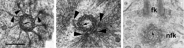
Structure of flagellar basal body in D. thienemanni (a, b) and non-flagellar basal body in M. ovata (c). a Transversal section of the FB distal end. Arrowheads show the prominent transitional fibers, connecting basal body to the plasma membrane. Note the arc-shaped filaments (arrow) inside basal body, interconnecting neighbor triplets. b Transversal section of FB middle part. Arrow shows the arc-shaped filaments inside FB; arrowheads point 4 separate MTOCs initiating the microtubular roots. c Transversal section of NFB (nfk). fk flagellar basal body. a, b after: [8]; c after: [11]. Scale bar 200 nm