
Fig. 1.
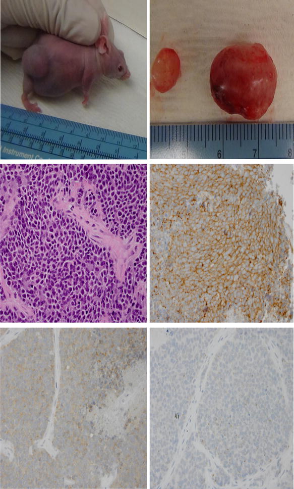
Subcutaneous growth of patient-derived xenograft in a SCID mouse host just prior to euthanasia. Harvested tumor from bilateral subcutaneous pockets; 3 × 3 mm sized sections were immediately propagated to the next generation of mice through implantation into subcutaneous pockets over the hind legs of the mice without in vitro manipulation (top panel). Histopathologic confirmation of small cell lung carcinoma histology by hematoxylin and eosin stain (X400) and immunohistochemistry for neuroendocrine differentiation showing intense diffusely positive staining for CD56 (middle panel), moderately intense staining for synaptophysin and focal areas of weakly positive chromogranin A staining (bottom panel)