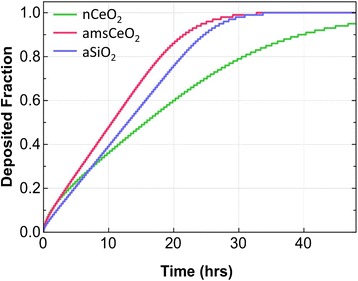
Fig. 1.

Fraction of deposited nCeO2, amsCeO2, and aSiO2 particles over a 48 h exposure, using fibroblasts cultured in 24-well tissue culture plates in fibroblast growth medium. The Harvard in vitro dosimetry method was used to determine the fraction of nanoparticle delivered to cells as a function of time. Uncoated nCeO2 settled slower than both amsCeO2 and aSiO2, with all nanoparticles achieving approximately 90 % deposition by 40 h post-administration to cells
