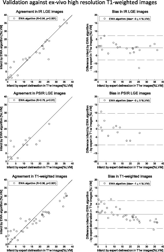
Fig. 4.

Validation against ex-vivo high resolution T1-weighted images: Scatter plots (left column) and Bland-Altman plots (right column) of infarct size expressed as % of left ventricular mass (%LVM) for the EWA algorithm against infarct size by expert delineation in ex-vivo high resolution T1-weighted images (T1w). Validation in in-vivo magnitude inversion recovery (IR, top row, n = 23 pigs), in-vivo phase sensitive inversion recovery (PSIR, middle row, n = 13) and ex-vivo high resolution T1-weighted images (T1w, bottom row, n = 38). Left column: solid line = line of identity; dashed line = regression line. Right column: solid line = mean bias; dashed line = mean ± two standard deviations
