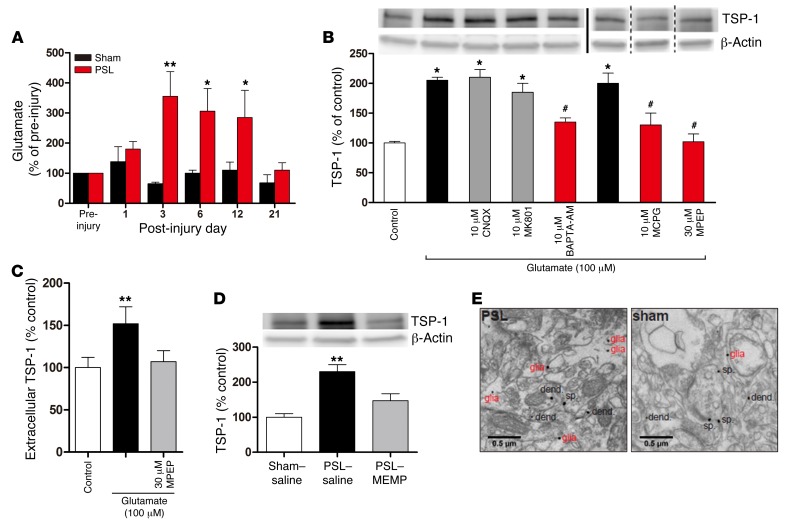
Figure 6. Increased extracellular glutamate and astrocytic mGluR5 signaling in S1 cortex following PSL injury mediates Ca2+-dependent TSP-1 release.
(A) Extracellular glutamate levels in S1 cortex transiently increased after PSL injury (n = 5 mice/group), but not after sham operation (n = 3 mice). *P < 0.05 and **P < 0.01 versus pre-injury, by 1-way ANOVA. (B) 100 μM glutamate-mediated increases in TSP-1 expression in cultured cortical astrocytes, as measured with Western blots, were inhibited by the mGluR5 antagonists MCPG and MPEP and by BAPTA-AM, but were unaffected by AMPA and NMDA antagonists (CNQX and MK-801). *P < 0.05 versus control; #P < 0.05 versus glutamate, by Kruskal-Wallis test. Note that the MCPG and MPEP data were run on separate gels, as indicated by the vertical lines. (C) Levels of extracellular TSP-1 released from cultured cortical astrocytes as measured by ELISA. Glutamate application significantly increased TSP-1 release, which was blocked by MPEP. **P < 0.01, by Kruskal-Wallis test. (D) Local MPEP infusion in vivo blocked the increase in TSP-1 levels in the S1 cortex following PSL injury. Representative Western blots demonstrated a significant increase in S1 cortex TSP-1 levels following PSL injury, which was inhibited by MPEP administration into S1 cortex (n = 4 mice/group). **P < 0.01, by 1-way ANOVA. Error bars in A–D represent the mean ± SEM. (E) Representative electron micrographs of contralateral (left) S1 cortex (layer I) labeled for mGluR5 in PSL-injured and sham control mice. Immunogold labeling for mGluR5 was found both in neuronal compartments (dend., dendrite; sp., spine) and glia, with the frequency of glia labeling increasing after PSL injury.

