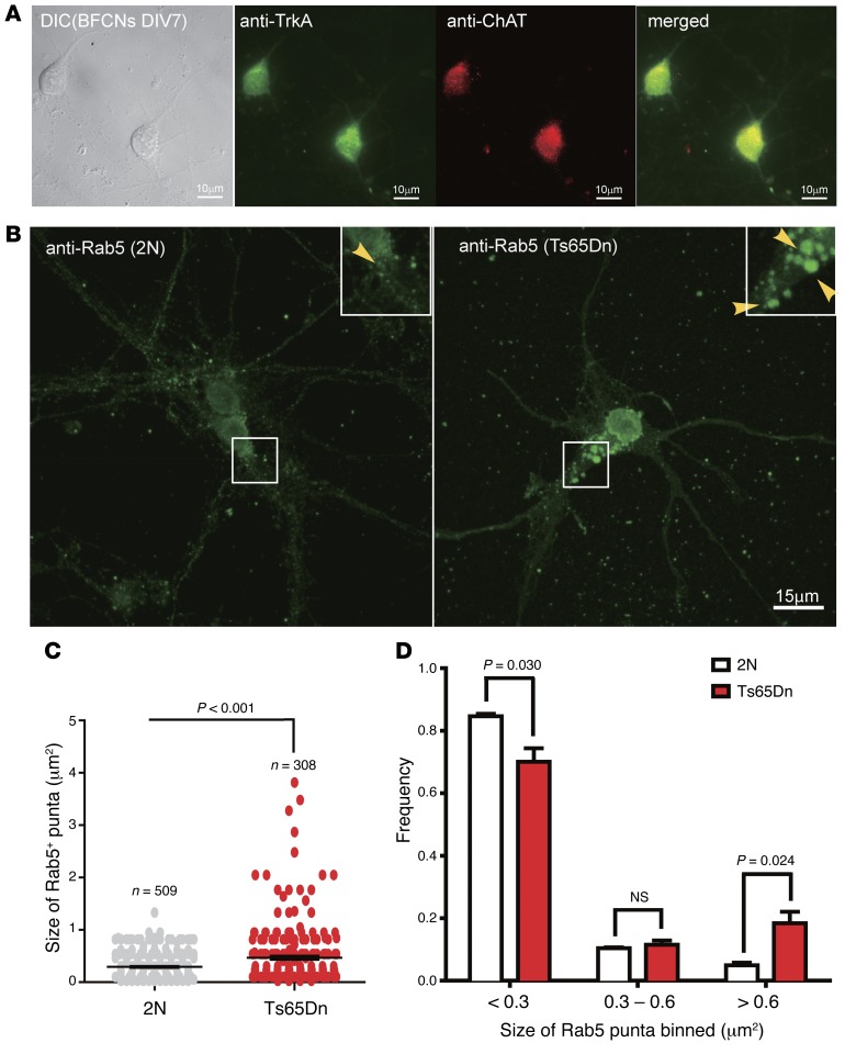
Figure 1. Rab5+ early endosomes were enlarged in primary BFCNs of Ts65Dn mice.
(A) Representative images of primary BFCNs (DIV7) were costained for the cholinergic neuronal marker ChAT (red) and the NGF receptor TrkA (green). DIC and merged images are also shown. Scale bars: 10 μm. (B) Representative images are shown for Rab5 staining of BFCNs from Ts65Dn (right) and 2N littermates (left). The sizes of Rab5+ puncta in BFCNs from Ts65Dn and 2N littermates were quantified using ImageJ. Insets: Zoom-in (×2) images of selected areas. Scale bar: 15 μm. (C) Measurement of Rab5+ puncta in B. The average area was 0.468 μm2 (n = 308) for Ts65Dn, 0.295 μm2 (n = 509) for 2N. The measurements were from 3 experiments, with 20–30 cells analyzed each time. (D) The size distribution of Rab5+ puncta in BFCNs from Ts65Dn mice showed a shift from smaller to larger binned areas in comparison to those from 2N littermates. All data represent mean ± SEM (n = 3), and P values were calculated using Student’s t test. n.s., nonsignificant.

