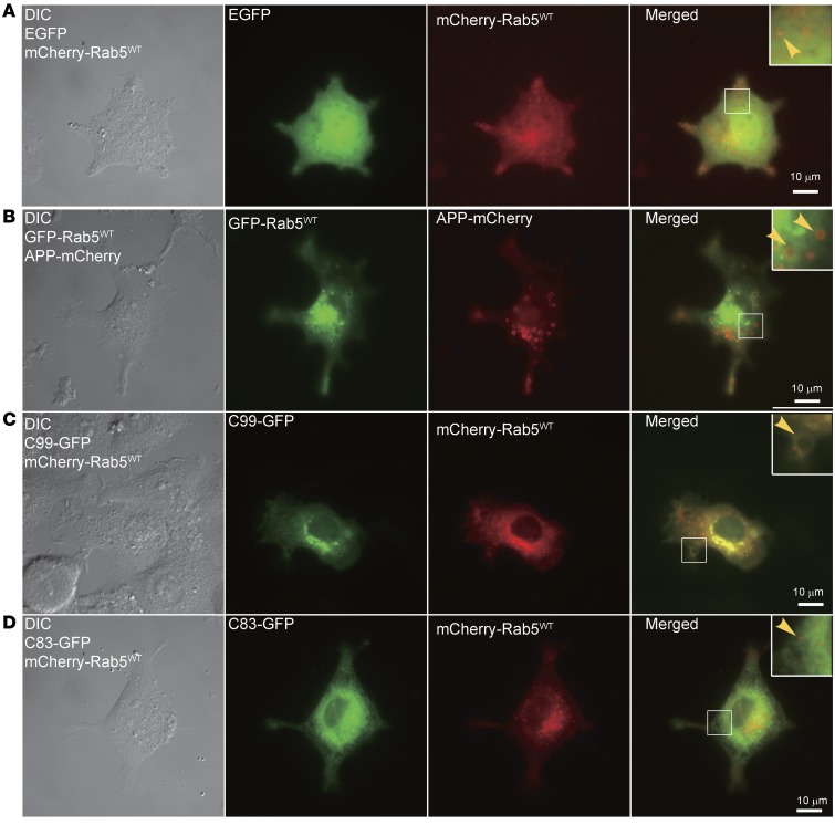
Figure 3. Full-length APP and C99, but not C83, induced the enlargement of Rab5+ endosomes in PC12M cells.
PC12M cells were cultured on glass coverslips and cotransfected with the indicated plasmids. Live cell imaging was performed as described in Methods. Images of DIC, FITC, and Texas Red channels were collected, and representative images are shown. Images for cotransfection of mCherry-Rab5WT with EGFP served as the control (A). (B) Cotransfection of EGFP-Rab5WT with APP-mCherry. Cotransfection of mCherry-Rab5WT with (C) C99-GFP or (D) C83-GFP. Insets: Zoom-in (×2.5) images of the selected areas. Scale bars: 10 μm.

