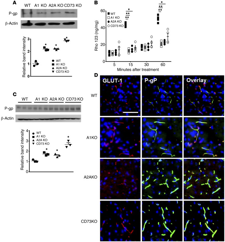
Figure 6. Ablation of CD73 or ARs increased P-gp expression and decreased P-gp substrate accumulation in the brain.
(A) Western blot analysis of P-gp from primary brain endothelial cells of brains of WT, A1, A2A, and CD73 KO mice. β-Actin was used as a loading control. Intensity of bands was normalized with that of β-actin. *P < 0.05 (n = 3 from 3 different Western blots, 2-tailed Student’s t test). (B) Rho123 uptake assay was performed using primary brain endothelial cells from brains of WT, A1, A2A, and CD73 KO mice. Cells were grown to confluence and treated with 2.5 μM of Rho123 at 5, 15, 30, and 60 minutes. Cells were lysed with lysis buffer and were analyzed by fluorometry, with excitation at 488 nm and emission at 523 nm. *P < 0.05; **P < 0.01 (n = 4, 2-tailed Student’s t test, 1 representative result of 2 different experiments). (C) Western blot analysis of P-gp expression levels from brains of WT, A1 KO, A2A KO, and CD73 KO mice. β-Actin was used as loading control. Band intensity was normalized to that of β-actin and graphed. *P < 0.05 (n = 3, 2-tailed Student’s t test). (D) IFA on the brain of WT, A1 KO, A2A KO, and CD73 KO mice. Frozen brain sections were stained with GLUT1 (an endothelial marker, red) and P-gp (green) and counterstained with DAPI (blue). Nucleus was counterstained with DAPI (blue). Scale bar: 100 μm.

