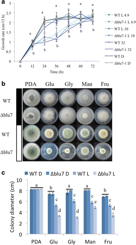
Fig. 4.

Reduced growth of the ∆blu7 mutant cultivated under constant light. a Colony growth rate of the ∆blu7 and WT strains registered every 12 h during 72 h when grown under exposure to 4.9, 10 or 32 μmolm−2s−1 blue-light on PDA. b Colonies of the WT and ∆blu7 strains grown on minimal media with glucose (Glu), glycerol (Gly), mannitol (Man) or fructose (Fru) as a sole carbon source, PDA was used as control. Colonies were photographed after 72 h of growth in the carbon source indicated, grown either in the dark (filled bar) or under illumination with white light of 3.2 μmolm−2s−1 (empty bar). c Colony diameters measured after 72 h of growth. One-way ANOVA and pairwise t-test was applied to the data. Different letters indicate statistically significant differences. Three independent biological replicates were used for each strain
