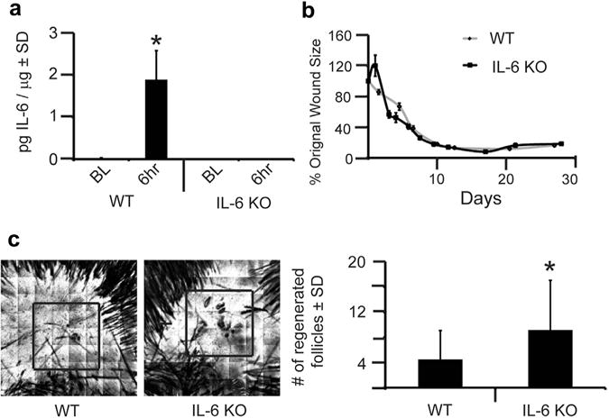
Figure 1. IL-6 knockout (KO) mice have increased wound-induced hair neogenesis (WIHN).

(a) Levels of IL-6 protein were measured by ELISA in wild-type (WT) and IL-6 KO mice, at baseline (BL) and 6 hours after wounding. n = 3. P < 0.05. (b) Full-excision skin wounding to the depth of skeletal muscle was performed in WT and IL-6 KO mice. At the time of wounding, we captured an image of the open wound. Subsequent images were captured every 1–2 days for 28 days. Using ImageJ computer software, wound size was calculated for each image, and data are shown as percentage of the original wound size (100%). Each datapoint is an average of 6–9 mice. (c) WIHN in IL-6 KO mice compared to WT control mice as measured by confocal scanning laser microscopy 20–24 days after wounding. Representative images for WT and IL-6 KO mice are shown. Area of WIHN is shown within box. Original image size is 4 mm2. n = 45. *P = 0.0004. SD, standard deviation.
