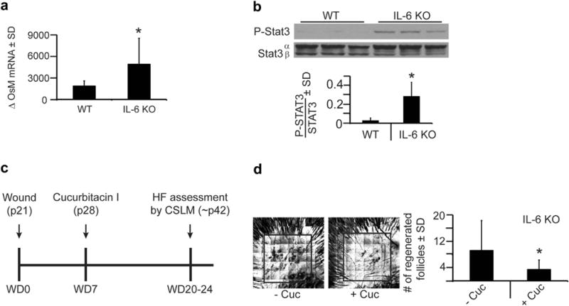
Figure 2. IL-6 knockout (KO) mice exhibit increased and necessary phosphorylated Stat3 (P-Stat3) signaling for wound-induced hair neogenesis.

(a) Quantitative real-time PCR of oncostatin M (OsM) levels 6 hours after wounding in wild-type (WT) and IL-6 KO mice. Shown as fold change relative to baseline. n = 5–6. *P = 0.01. (b) Levels of P-Stat3 protein expression in WT and IL-6 KO mice in normal, nonwounded skin (baseline) as measured by western blot. Bar graph depicts ratio of P-Stat3/Stat 3 in arbitrary units as measured by Image J software. n = 3. *P = 0.02. (c) Full-excision skin wounding to the depth of skeletal muscle performed in WT and IL-6 KO mice, as in Figure 1c, and 7 days after wounding (WD7), cucurbitacin I (2 mg/kg), or control (phosphate buffered saline) was injected into the healing wound. Hair follicles (HF) were assessed by CSLM ~ 2 weeks later. (d) Number of regenerated hair follicles within the scar, as assessed in Figure 1c, in IL-6 KO mice with cucurbitacin I (+Cuc) compared to vehicle (−Cuc). Representative confocal scanning laser microscopy images are shown. Area of wound-induced hair neogenesis shown within box. Original image size is 4 mm2. n = 11–13. *P < 0.05. SD, standard deviation.
