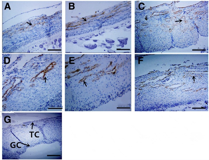
Figure 4.
Immunohistochemistry detection of chicken MMP13 protein in different follicles from the 159-d-old hen ovaries. (A) SW, (B) SY, (C) F5, (D) F3, (E) F1, (F) POF1, and (G) negative control with PBS used in place of the primary antibody. Arrowheads indicate the position of strongly stained protein. GC, granulosa cells; TC, theca cells. Bar for (A–G): 200 μm

