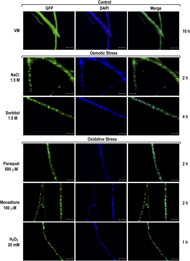
Figure 4.
SEB-1 protein translocates to the nucleus under osmotic and oxidative stress conditions. Conidia from the Δseb-1 complemented strain (Δseb-1 his-3::Pccg-1-seb1-sfgfp) were grown on coverslips in liquid VM medium containing 1% sucrose at 30° for 10 hr. After this period, mycelia were subjected to osmotic and oxidative stresses for 1 hr, 2 hr, or 4 hr. Stressed mycelia were fixed in PBS with formaldehyde, the nuclei were stained with DAPI, and the fluorescence were visualized. Mycelia from liquid VM medium cultured for 10 hr at 30° was used as control. Fluorescence was evaluated using the AXIO Imager.A2 microscope (Zeiss) at a magnification of 100×. The images are representative of at least two independent experiments. Scale bar: 10 μm. DAPI, 4’,6-diamino-2-phenylindole; GFP, green fluorescent protein; PBS, phosphate-buffered saline; VM, Vogel’s minimal.

