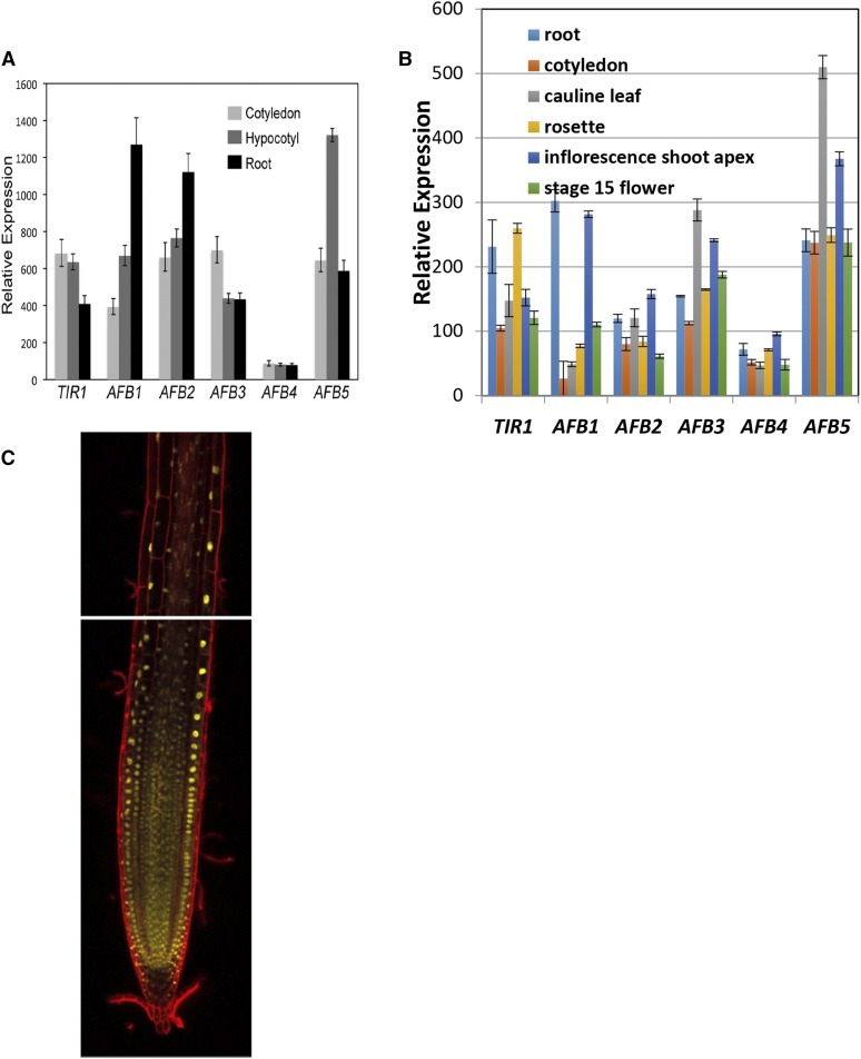
Figure 7.
Expression of the AFB4 and AFB5 genes. (A) qRT-PCR of TIR1/AFB genes in 4-day-old WT seedling tissues grown under SD conditions. Primer pairs are listed in Table 1 with AFB4-4 and AFB5-4 being used for those respective genes. Expression is normalized to PP2AA3 using the PP2AA3-S primer pair. Error bars represent standard error. (B) TIR1/AFB expression levels in various tissues. Replotted from Winter et al. (2007). (C) mCitrine fluorescence (yellow) was visualized in roots of the AFB5-mCitrine line #9 using confocal microscopy. Cells were stained with propidium iodide (red).

