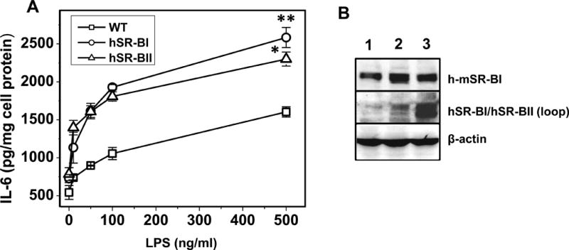
Figure 10. Dose-dependent LPS-induced IL-6 secretion in primary kidney epithelial cells (KECs) from WT, hSR-BI and hSR-BII transgenic mice.

A. KECs were incubated with LPS (0, 10, 50, 100 and 500 ng/ml) in serum-free DMEM containing 2mg/ml BSA and 10mM HEPES pH 7.4 for 18 hours. Media were collected and assayed for IL-6 secretion by ELISA.
* P<0.05, ** P<0.01 vs WT LPS-treated cells. B. Western blot analysis of KECs from WT (lane 1), hSR-BI tgn (lane 2) and hSR-BII tgn (lane 3) mice. The expression of hSR-BI and mSR-BI expression was detected utilizing an anti-SR-BI antibody (against C-terminal 450–509 amino acid peptide, Novus Biologicals, cat. # NB400-101) and hSR-BI/hSR-BII protein was detected using anti-human SR-BI/BII loop antibody (against 104–294 amino acid peptide, BD Biosciences). Protein expression of β-actin was measured as the loading control.
