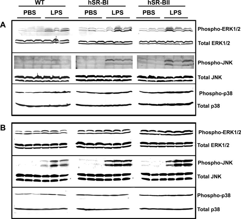
Figure 9. Western Blot analyses of MAPKs activity in the liver and kidney of LPS-treated WT and SR-B transgenic mice.

Four hours after PBS or LPS (1 mg/kg, IP) injection into WT, hSR-BI and hSR-BII tgn mice (n=3 for each group), mice were sacrificed; organs were collected and processed for assessment of MAPKs phosphorylation as described in the Materials and Methods section. A. MAPK activity of liver samples using antibodies against phospho-ERK1/2 (upper panel) or phospho-p38 (bottom panel). B. MAPK activity of kidney samples using antibodies against phospho-ERK1/2 (top panel), phospho-JNK (middle panel), or phospho-p38 (bottom panel). Equal loading of samples was ensured by using anti-ERK1/2, anti-JNK or anti-p38 MAPK antibodies against total (non-phosphorylated) MAPK protein.
