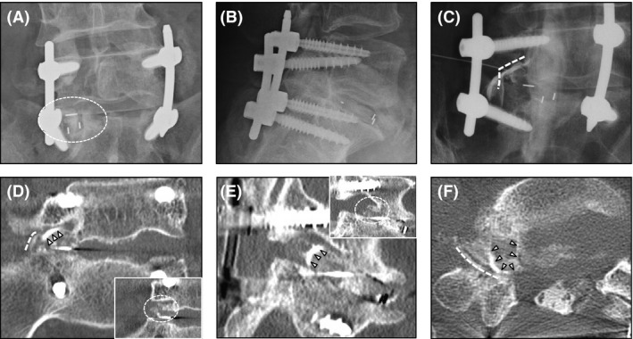
Figure 2.

Radiological examinations 3 months after the primary surgery with right L5 radiculopathy. (A) Marked subsidence of the intervertebral cage is shown (dotted circle). (B) lateral X‐ray did not suggest apparent back‐out of the cage, nor subsidence to the S1 endplate. (C) Spinal nerve enhancement was performed just after the myelography. There was a rectangular‐shaped rim enhancement which knocked up the spinal nerve (dotted line). (D–F) Computed tomographic (CT) myelography. Enhanced rim of the cage (arrowheads) showed knocking up of spinal nerve (dotted line) at the foramen. Also, there was an obscure bone‐density mass in the foramen in the plain CT image (small images in (D) and (E)), indicating marginal fracture (dotted circle). Laterally installed intervertebral cage was considered to be knocking up the L5 spinal nerve with marginal fracture.
