Figure 1.
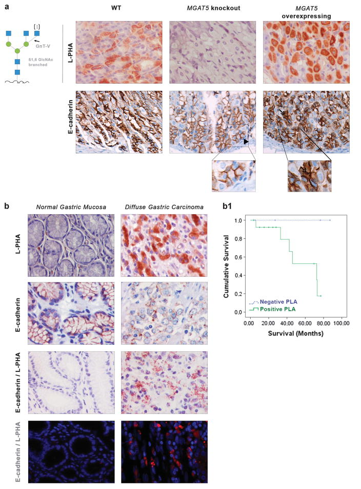
Evaluation of the expression of E-cadherin and β1,6 GlcNAc-branched structures in the gastric mucosa of MGAT5 knockout and MGAT5 overexpressing and in human normal gastric mucosa versus gastric carcinoma. (a) L-PHA histochemistry detecting the β1,6 GlcNAc-branched N-glycans catalyzed by GnT-V showed a moderate expression of the β1,6-branched structures in the gastric mucosa of WT mice. No positive L-PHA reactivity was observed in the gastric mucosa of MGAT5 KO mice, whereas a clear overexpression of β1,6 GlcNAc-branched structures was detected in MGAT5 transgenic gastric mucosa. MGAT5 KO mice showed a normal E-cadherin expression at the basolateral cell surface (arrowhead). In MGAT5 transgenic mice, immunoexpression of E-cadherin was displayed aberrantly in the cytoplasm of mucous neck cells (not shown) and in deep glands of the gastric mucosa (arrow). (b) In situ PLA showed weak/absence PLA signal in normal gastric mucosa and a marked positivity of PLA signal (brown dots for brightfield and red dots for immunofluorescence) in neoplastic cells from gastric carcinoma, demonstrating a profound modification of E-cadherin with the β1,6 GlcNAc-branched structures in human gastric cancer (×40, original magnification). (b1) Survival rates of patients with diffuse gastric cancer (DGC) accordingly with PLA signal (positive versus negative). Kaplan–Meier curves demonstrate the probability of overall survival for patients with DGC accordingly with the presence/absence of GnT-V-mediated aberrant glycosylation of E-cadherin (P = 0.093).

