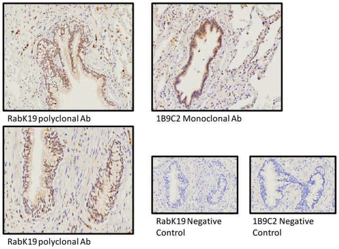
Fig 2. Immunohistochemical analysis showed that MAP3K19 expression in normal lung is predominantly limited to bronchial epithelial cells and interstitial and alveolar macrophage.
Normal human lung was stained with either the 1B9C2 anti-MAP3K19 mouse monoclonal antibody (brown staining) or RabK19 rabbit polyclonal antibody (brown staining) and counter-stained with hematoxylin.

