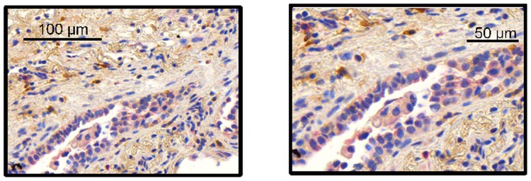
Fig 5. MAP3K19 is expressed in the atypical epithelium tissue that lies adjacent to the fibroblastic foci.
MAP3K19 staining of a lung biopsy from a rapid IPF progressor patient showed clear, albeit lower intensity staining, of MAP3K19 (red staining, RabK19 Ab) in the atypical epithelium adjacent to the fibroblastic foci. This staining pattern of the atypical epithelium was found in multiple biopsy sections from IPF patients (n = 3). This section was counter-stained with SSEA-4 (brown stain).

