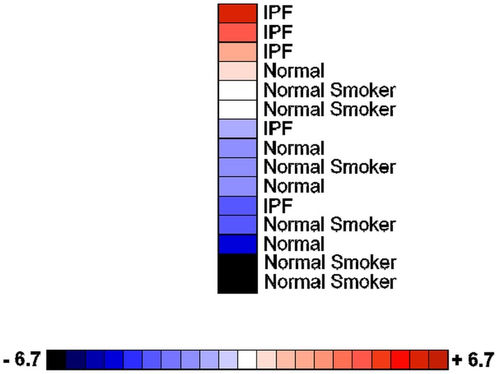
Fig 7. MAP3K19 is over-expressed in bronchoalveolar lavage macrophages from IPF patients.
Heatmap showing MAP3K19 expression in BAL macrophages isolated from mild-to-moderate IPF patients (n = 5), normal controls (n = 4) and healthy smokers (n = 6) as determined by RT-qPCR analysis. The data are normalized to GAPDH expression and the fold changes are depicted on the color scale. The RT-qPCR is representative of two independent experiments (biological replicates) performed in duplicate. Anova analysis of MAP3K19 expression levels among the different groups revealed that the difference in mRNA levels between the IPF group, with the lowest expressing patient omitted, and either the normal patients or smokers was significant, P<0.01.

