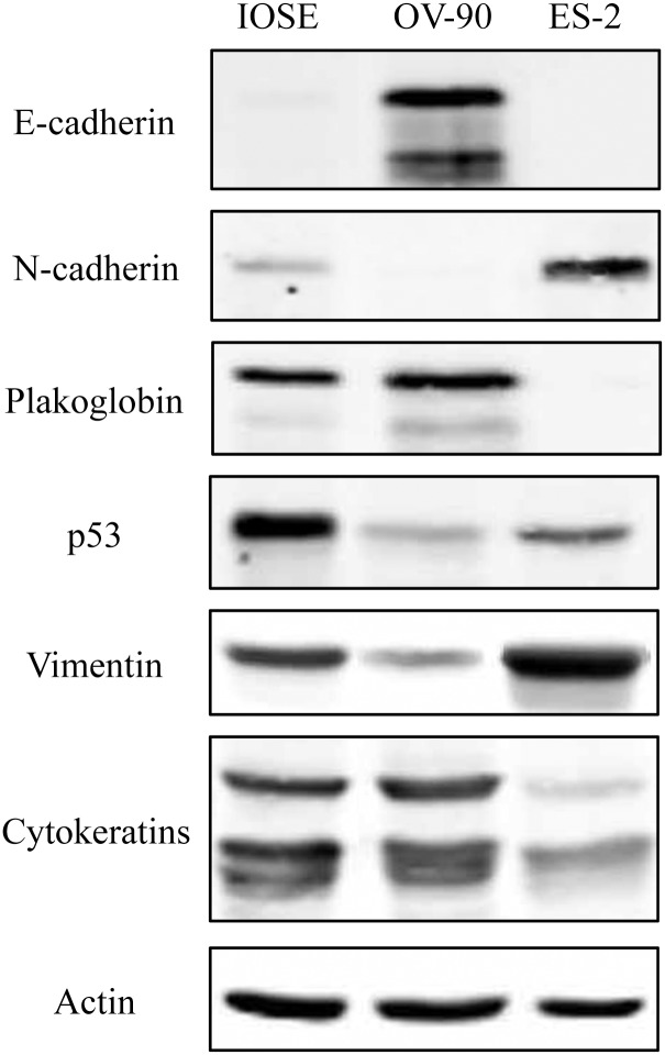
Fig 1. Protein expression of epithelial and mesenchymal markers and p53 in OVCA cell lines.
Total cell lysates from IOSE-364, ES-2 and OV-90 cells were processed for immunoblot analysis using N-cadherin, E-cadherin, plakoglobin, vimentin, cytokeratins and p53 antibodies. Equal loadings were confirmed by processing the same lysates with actin antibodies.

