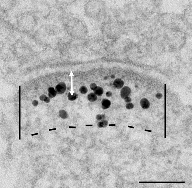
Fig 1. Electron micrograph of an asymmetric synapse from dissociated hippocampal culture labeled for Shank3 with ab2.

Labeling intensity at the PSD is measured by counting all particles within the marked area 120 nm below the postsynaptic membrane, divided by the length of the PSD, and expressed as number of labels/μm PSD. Distance of label from the postsynaptic membrane is measured from the center of the particle to the outer edge of the membrane (white arrow). Scale bar = 0.1 μm.
