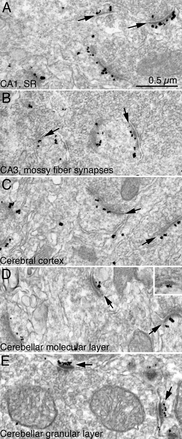
Fig 3.
Electron micrographs from perfusion fixed-mouse brain showing immunogold labeling for Shank3 using ab1 (A-E) and ab3 (D, inset). PSDs (arrows) were labeled in all regions examined, including hippocampus [stratum radiatum (SR) of the CA1 region (A), mossy fiber synapses in the CA3 region (B)], cerebral cortex (C), and the outer molecular layer (D) and granular layer (E) of the cerebellum. Scale bar = 0.5 μm.

