Fig 5.
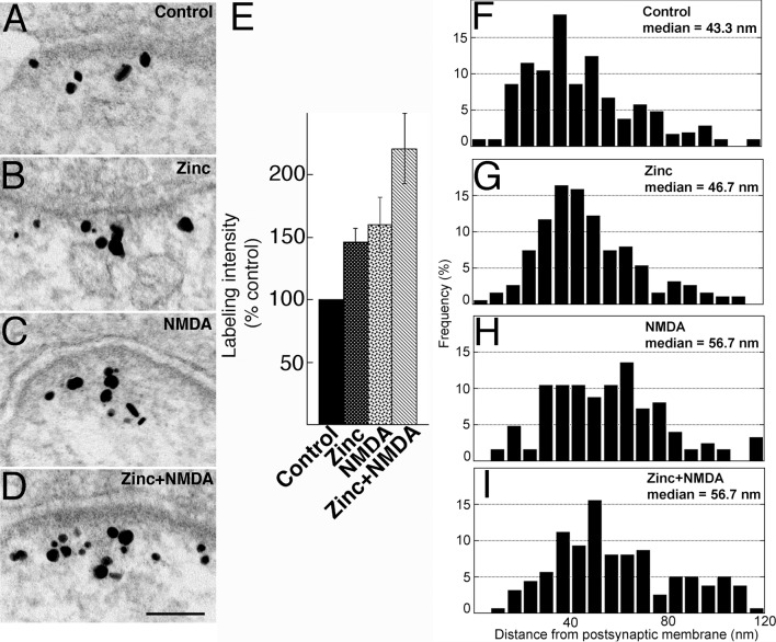
(A-D) Electron micrographs of asymmetric synapses labeled for Shank3 using ab1. Hippocampal cultures were pre-incubated for 1 hr in media with or without zinc and subsequently treated for another 2 min in the same media with or without NMDA, as indicated. After 1 hr zinc incubation (B) or 2 min NMDA treatment (C), labeling intensities were higher than that of control (A). Upon pre-incubation with zinc followed by NMDA treatment (D), labeling intensity was even higher than after either treatment alone. Scale bar = 0.1 μm. (E) Bar graphs represent combined data (mean ± SEM) from S2 Table. (F-I) Median distance of gold particles from the postsynaptic membrane increased upon NMDA treatment, either in the absence (H) or presence of zinc (I), but not in samples incubated with zinc only (G) when compared to control (F).

