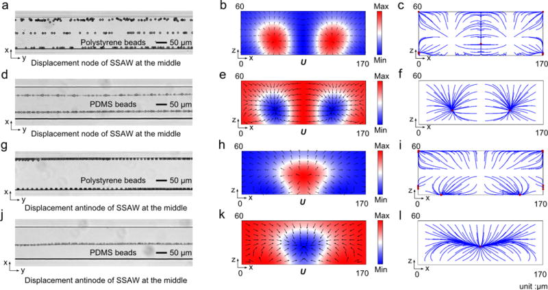
Fig. 2.

Microparticle acoustophoresis in PDMS channels with a width of 170 μm. (a)–(c), Particle traces and numerical results for polystyrene beads when the displacement node is located in the middle of the channel. (d)–(f), Particle traces and numerical results for PDMS beads when the displacement node is located in the middle of the channel. (g)–(i), Particle traces and numerical results for polystyrene beads when the displacement antinode is located in the middle of the channel. (j)–(l), Particle traces and numerical results for PDMS beads when the displacement antinode is located in the middle of the channel. (a), (d), (g), and (j), experimental particle traces in the x-y plane under the mentioned conditions (a, g: polystyrene beads; d, j: PDMS beads). (b), (e), (h), and (k), Numerical results of radiation force potential and acoustic radiation forces in the x-z plane for the mentioned cases (b, h: polystyrene beads; e, k: PDMS beads). (c), (f), (i), and (l), Numerical results of bead trajectories and final locations in the x-z plane for the mentioned cases (c, i: polystyrene beads; f, l: PDMS beads).
