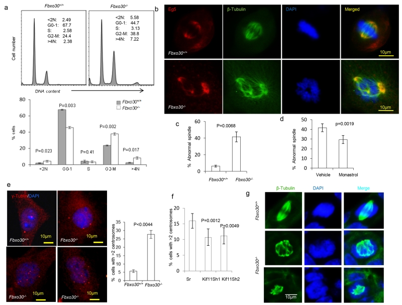
Fig. 7.
Fbxo30 deletion causes chromosomal instability and spindle defects in vivo and in vitro. a. DNA content analysis based on propidium iodide staining of ethanol-fixed MGE cells after 2 passages in vitro. Representative FACS profiles are shown on top and the summary data involving 3 mouse samples per group shown in the bottom. Note a major increase in cells with > 4N in DNA content. b. Deletion of Fbxo30 causes formation of multipolar mitotic spindles. WT (upper panels) and Fbxo30−/− (lower panels) MGE were stained for EG5, tubulin, and DNA. c. Summary data for three different cultures (n=3). d. Monastrol treatment reduces % of MGE cells with abnormal spindles. As in c, except that the MGE cells prepared from Fbxo30−/− mice were treated with either vehicle control or monastrol (30 nM) for 48 hours prior to staining. e. Fbxo30 deletion causes abnormal centrosome amplification. The images on the left depict γ-tubulin distribution in a WT and 3 Fbxo30−/− MGE cells, while the right panel shows the mean and SEM of percent cells with greater than 2 centrosomes (n=15) and has been repeated 3 times. f. ShRNA silencing of the Kif11 gene restores centrosome homeostasis in Fbxo30−/−MGE cells. Fbxo30−/− MGE cells were cultured in the presence of either vehicle or a low dose of monastrol (30 nM) for 48 hours. The centrosome number was determined by γ-tubulin staining. Data shown are means and SEM of percent cells with >2 centrosomes (n=10) and have been repeated 3 times). g. Accumulation of multi-polar spindle mitotic cells in the Fbxo30−/− mammary glands, as revealed by staining with anti-β-tubulin mAb and DAPI staining of frozen mammary gland sections. The top panels show a cell with a normal bipoplar spindle in WT mammary gland; the lower panels show a cell in Fbxo30−/− mammary gland with multipolar mitotic spindle and abnormal distribution of chromatin. Cells with mitotic spindle are difficult to find in vivo. Only 1 such cell was found in five slides prepared from 5 virgin mice, while more than 50 cells with mitotic spindle were found in 2 of the 5 slides from the 5 virgin Fbx30−/− mice. None of the mitotic spindles have normal morphology. Seven such cells are shown in supplemental Fig. S3.

