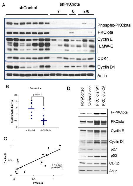
Figure 2. Downregulation of PKCiota decreases the expression cyclin E.
(A) shControl (CCTAAGGTTAAGTCGCCCTCGCTCGAGCGAGGGCGACTTAACCTTAGG) and shPKCiota clones (constructs 7, 8, and 7/8) (construct 7-sequence TGACCAGAACACAGAGGATTATCTCTTCC targeting exon 14; construct 8 CAGGAGATACAACCAGCACTTTCTGTGGT targeting exon 13), of IGROV cells were subjected to Western blot analysis using phospho-PKCiota, PKCiota, cyclin E, CDK4, cyclin D1, and actin (as loading control). (B) Western blots from panel A were subjected to densitometric analysis and the resultant values for cyclin E (full length and LMW-E) were plotted for each of the shControl and shPKCiota clones. p=0.0021 (C) Linear regression of cyclin EL densitometric values as a function of PKCiota for each of the clones in panel A were plotted. (r=0.803; p= 0.0005). (D) IGROV ovarian cancer cells were transfected with shControl, shPKCiota PKCiota (Wt-wildtype, Ca-constitutively active (A129E), or DN-dominant negative (K122STOP), and GFP (negative control) and harvested at 24 and 48 hours post transfection and subjected to cell sorting for GFP positive cells. Immediately following cell sorting the GFP+ cells were lysed and subjected to western blot analysis using antibodies to phospho-PKCiota, PKCiota, cyclin E, cyclin D1, CDK2, and p53. Actin was used as a loading control.

