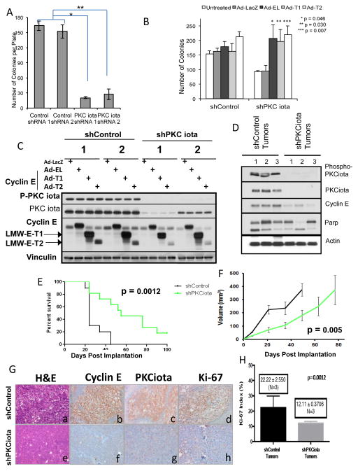
Figure 5. PKCiota knockdown inhibits soft agar colony and tumor formation through inhibition of cyclin E.
(A) The number of colonies formed in soft agar for shControl and shPKCiota clones after 15 days was determined. * p = 2.7 ×10⁻⁷; ** p = 4.4 × 10⁻⁵. (B) IGROV shControl and shPKCiota stable clones were infected with Ad-LacZ, Ad-Cyclin EL, Ad-LMW-E-T1 or –T2 and were plated in soft agar for 10 days at which point the colonies were enumerated. Number of colonies were statistically different between LacZ, shControl versus shPKCiota (p = 0.001). (C) Western blot analysis confirming the infection of the IGROV stable shRNA clones with Ad-cyclin E. Phospho-PKCiota and PKCiota antibodies. Vinculin was used as a loading control. (D–H) 1 × 10⁶ shControl IGROV and shPKCiota IGROV cells were injected into 2 groups of 5 nude mice and tumor volume was monitored over time. Mice were euthanized when tumor volume reached 1.5 cm. The experiment was repeated 3 times, each time with two groups of 5 mice in each injection arm. (D) Western blots were performed on the tumor tissue dissected from mice, upon sacrifice to assess PKCiota, phospho-PKCiota, cyclin E, and cleaved Parp expression. Actin was used as a loading control. (E) Kaplan-Meier estimates of surviving mice (15 in each group) was generated. (F) Tumor volume of mice (15 in each group) are plotted (G) Representative sections of invasive breast tumors from shControl and shPKCiota examined histology by H & E (a, e) expression of cyclin E (b,f), PKCι (c,g) and Ki-67 (d,h) antibodies. Representative section are at ×200 magnification H) Ki67 index was expressed as percentage of positive cells in 600 tumor cells. N=3 for each condition (tumors from ShControl and shPKCiota mice).

