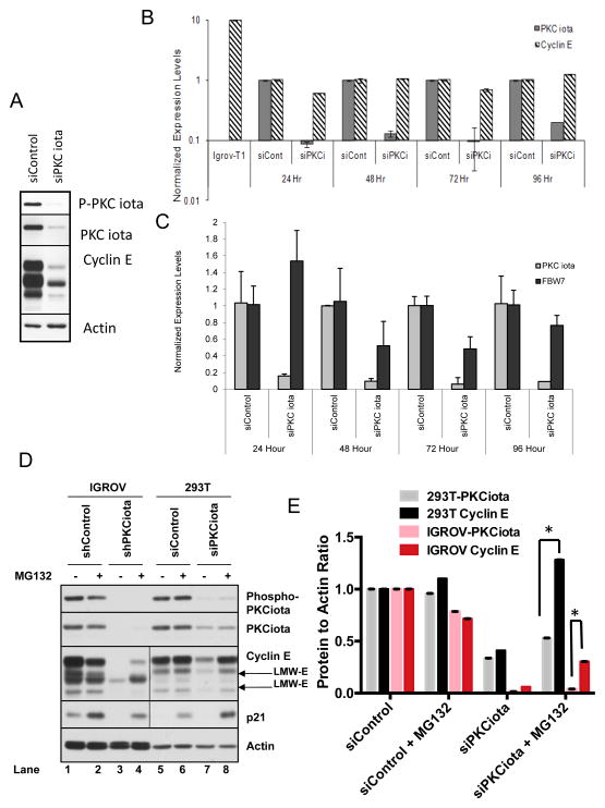
Figure 6. PKCiota regulates cyclin E through its degradation rather than transcription.
IGROV cells were transfected with siControl or siPKCiota and subjected to (A) western blot analysis with the indicated antibodies and (B, C) qRT-PCR. To this end upon transient knockdown of PKCiota for 24, 48, 72 and 96 hours RNA was harvested at each time point and used for measurement of (B)cyclin E and PKCiota levels or (C) total FBW-7 levels. IGROV cells infected with cyclin E-T1 (IGROV-T1) was used as control for cyclin E primers. GAPDH was used as a loading control in the qRT-PCR assay and the results for cyclin E and PKCiota for each condition was normalized with GAPDH. Experiment was repeated 3 times. (D) IGROV stable shPKCiota cells and 293T transient siPKCiota cells were treated with MG132, and western blot analysis was performed using phospho-PKCiota, PKCiota, cyclin E, p21 (control for MG132 response), and actin (loading control). Although all samples were run on the same gel, the exposure time for cyclin E and p21 Western blots were much longer in the 293T cells than the IGROV cells due to the low endogenous levels of these two proteins in 293T cells. (E) Cyclin E and PKCiota levels were quantitated by densitometric analysis for (D) 293 T and IGROV cells.

