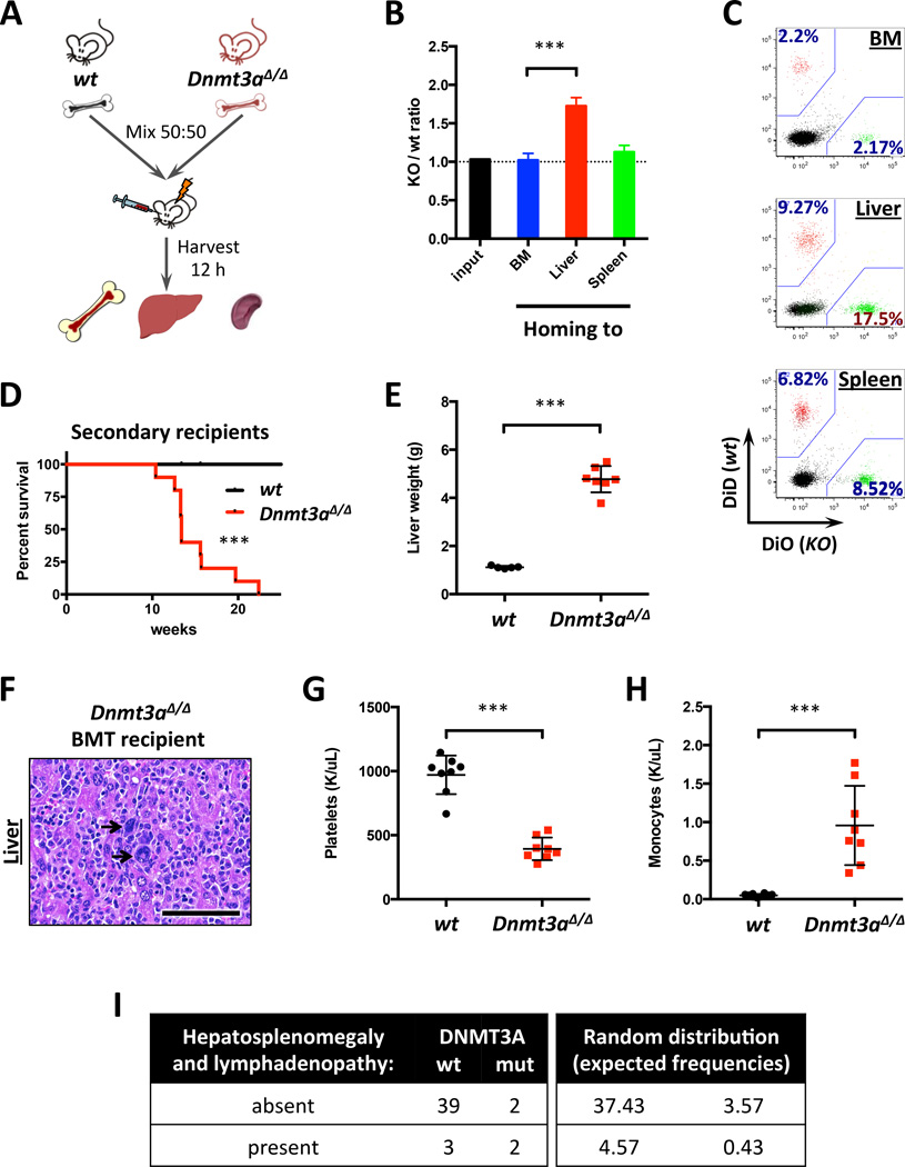
Figure 5. Conditional loss of Dnmt3a results in cell-autonomous liver tropism.
A. Schematic depiction of the homing assay workflow.
B. Relative homing tropism calculated as Dnmt3a KO/wt ratio in the bone marrow, livers and spleens of recipient mice (n=4; ***, p<0.001). Dotted line represents ratio at time of injection (1.0).
C. Representative FACS plots showing distribution of fluorescently labeled WT and Dnmt3a KO cells homed to the indicated sites, gated on CD45+.
D. Survival of lethally-irradiated recipient mice transplanted with total bone marrow from diseased Dnmt3a-null or control animals (n=10; ***, p=0.0003 by Gehan-Breslow-Wilcoxon test).
E–F. Liver weights (E; n=5–7; ***, p<0.001) and H/E-stained sections (F) in primary recipients transplanted with Dnmt3a-ablated bone marrow cells at time of disease onset and in control wild-type mice. Arrows – megakaryocytes indicative of EMH.
G–H. Platelet (G) and monocyte (H) counts in the peripheral blood of mice transplanted with Dnmt3a-null bone marrow (n=8; ***, p<0.001).
I. Increased frequency of extramedullary hematopoiesis in CMML patients with DNMT3A mutations. Left panel represents observed frequencies of hepatosplenomegaly and lymphadenopathy in patients with different DNMT3A (wt or mutant) status (n=46); right panel shows expected frequencies for random distribution (p=0.053, Phi=+0.39, 2-tailed Fisher’s exact test).

