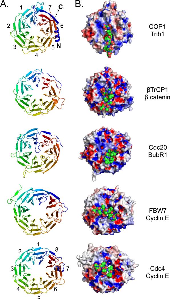
Figure 3. Comparison of WD40 β-propellers from E3 ligases bound to peptide motifs.
A. Cartoon representation, showing a top view (clockwise from N- to C-terminus) of seven bladed WD40 β-propellers from COP1, β-TRCP1 (1P22), CDC20 (4GGA) and eight bladed propellers from CDC4 (1NEX), and FBW7 (2OVQ). B. Surface representation of these propeller domains bound to cognate peptides represented as space-filling spheres. The propeller domains are colored according to electrostatic surface potential, and the bound substrates are shown in CPK colors (carbon: green).

