
Fig. 4.
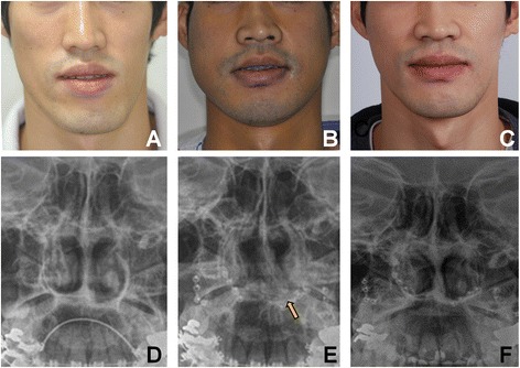
Clinical and radiographic findings of the case 3 patient before Le Fort I osteotomy (a, d), immediate after Le Fort I osteotomy (b, e), The arrow indicates the caudal part of the septal cartilage (e). Sepal deviation was successfully resolved after the corrective surgery (c, f)