Fig. 4.
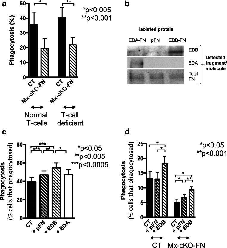
T-cells do not affect phagocytosis in the absence of fibronectin. a Independent of whether immune cells produced fibronectin or not, the absence of T-cells did not measurably affect phagocytosis. N = 2–5 mice per measurement and six measurements/group. EDB-containing fibronectin enhances phagocytosis. b The presence of the EDB domain, but not the EDA domain was confirmed in isolated EDB fibronectin (FN) by western blotting. Total fibronectin was detected in the three isolated isoforms. Same amount of fibronectin was added in the wells of each gel and blotted with either an antibody directed against EDA, against EDB or against total fibronectin as shown on the right. In the gel for total fibronectin, the amount of fibronectin added was 4-fold less than for the isoforms. c Adding control fibronectin (plasma fibronectin: pFN) slightly increased phagocytosis in human PMNs, but adding EDB fibronectin prior to adding S. aureus enhances phagocytosis. EDA fibronectin failed to enhance phagocytosis. Cells were isolated from four healthy subjects and treated with plasma-opsonized S. aureus. Prior to adding the bacteria to the cells, pFN or EDB fibronectin was added at a concentration of 200 ng/ml, which is 5-fold the concentration of EDB fibronectin in CSF. Phagocytosis measured after incubating the cells for 10 min at 37 °C and compared to cells treated similarly but left at 4 °C instead. N = 13 replicates except for EDA: five replicates. d Cells from control (CT) mice are not affected by adding pFN, but EDB fibronectin enhances phagocytosis. Mx-cKO-FN mice show enhanced phagocytosis already by addition of pFN but EDB fibronectin effect is more pronounced. Cells were similarly isolated and treated for 45 min. N = 7 pools of blood from four to six mice each/pooled group

