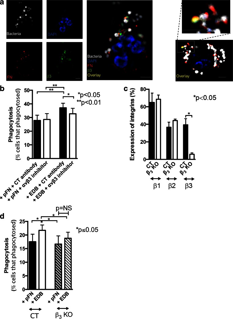
Fig. 5.
EDB fibronectin enhances phagocytosis through activating β3 integrin. a EDB fibronectin (FN) in red, β3 integrin in green, and bacteria in white are found in the proximity of each other. DAPI (blue) was used as a nuclear stain. PMNs were exposed to opsonized bacteria for 30 s, followed by fixation and staining. On the left, details of one cell are shown. On the right, a second example with a higher magnification of a clump of bacteria with fibronectin and β3 integrin co-staining is shown. N = 3 experiments. Bars represent 2.5 μm. b Using an αvβ3 inhibitory antibody results in diminished EDB-mediated phagocytosis (concentration used, 10 μg/ml). N = 8. c In β3 knockout mice, the percentage of PMNs expressing β1 or β2 integrin is similar, but β3 is deleted. d In the absence of β3, phagocytosis is no longer enhanced in the presence of EDB fibronectin (n = 5 pairs)

