Figure 5.
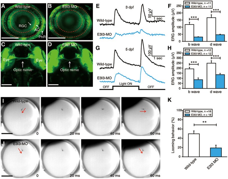
Electrophysiological recordings and behavioral tests showed that VIT is necessary for normal physiological function of zebrafish retina. (A, B) Confocal images showing GCaMP1.6 expression in photoreceptors (PhR), retinal ganglion cells (RGC) and amacrine cells (AC) of a 3-dpf Tg (Ath5-gal4:UAS-GCaMP1.6) larva (A) and a VIT morphant (B). The two red dash circles indicate the position of inner and outer plexiform layers. Scale bar, 100 μm. (C, D) Confocal images showing the projection of optic nerves from two eyes of a 3-dpf wild-type larva (C) and a VIT morphant (D). Scale bar, 100 μm. (E, G) ERG recording averaged from ten responses to a 2-s light ON stimulus in 3-dpf (E) or 5-dpf (G) wild-type larvae and VIT morphant. (F, H) Summary of ERG recording data at 3 (F) or 5 dpf (H). (I, J) Examples showing a looming stimulus-induced escape behavior in a 5-dpf wild-type larva (I), but not in a 5-dpf VIT morphant (J). The red arrows indicate the body axis of the larvae before and after looming-induced C-startle escape. Scale bar, 1 cm. (K) Summary of data showing the probability of looming-induced escape behaviors in 5-dpf wild-type larvae and VIT morphant. The number in the inset of F, H and K represents the number of larvae examined. **P < 0.01, ***P < 0.001; two-tailed unpaired Student's t-test for the data in F, H and K. Data are represented as mean ± SEM.

